Visualize the size, depth and location of vessels and surrounding anatomy as part of patient assessment and access.


- Overview
- Products & Accessories
- EIFU & Resources
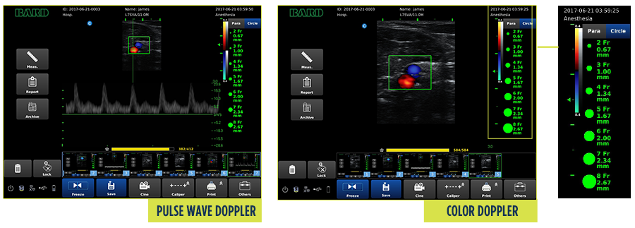
Probe options and application presets quickly apply image settings to the anatomy being assessed using application presets.
Assess the size, depth, and location of vessels as well as the surrounding anatomy.
Compare vessel to on-screen catheter icons to help assess the catheter-to-vein ratio.
Distinguish veins from arteries with Color Flow or Pulse Wave Doppler.
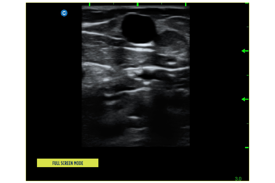
Use ultrasound guidance to help reduce needle passes, needle redirects and help increase first stick success rates.
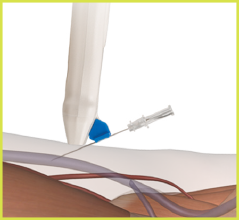
Needle guides
Compatible with Site-Rite™ and Pinpoint™ GT needle guides*.
Guides the needle to a designated depth under the ultrasound beam.
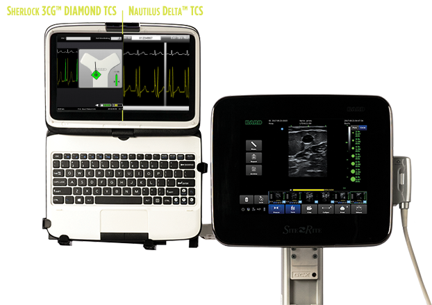
Using Stand-alone Tip Confirmation Systems (TCS) eliminate chest X-ray confirmation of catheter tip location for a range of devices and patients by configuring your system with the stand-alone Sherlock 3CG+™ TCS.
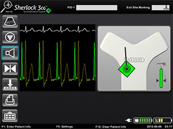
Sherlock 3CG+™ TCS
ECG tip confirmation for patients with a present, identifiable and consistent P-wave when placing CVADs.
Sherlock 3CG+™ TCS may be used for
- Adults and adolescents, 12–21 years, for the insertion of PICCs, CVCs, implantable ports and hemodialysis catheters
- Children, infants and neonates, for the insertion of PICCs and CICCs
Please consult product labels and inserts for indications, contraindications, hazards, warnings, precautions and directions for use.
*Pitts S, Ostroff M. The Use of Visualization Technology for the Insertion of Peripheral Intravneous Catheters. AVA Position Paper. 2021. Pg 2.
BD-66803 (7/22)

