true
Icon to close the modal
Support
Sales
Modal Close Icon
Icon to close thank you modal
Thank you for contacting our sales team!
A sales representive will get in touch with you shortly.
Customer Care

Sherlock 3CG™ Diamond Tip Confirmation System

Getting to the heart of the matter

Contact Us
sherlock_3cg_diamond_hero v2
Loading
Overview

Highlights

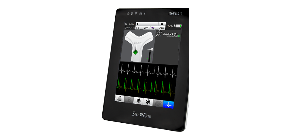
The Sherlock 3CG™ Diamond Tip Confirmation System (TCS) fully integrates magnetic tracking and ECG-based peripherally inserted central catheter (PICC) tip confirmation technology, which includes P-wave highlighting and max P-wave identification in adult patients with a present, identifiable, and consistent P-wave.
true
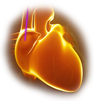
3cg-heart.png
Getting to the Heart of the Matter

Clinicians using Sherlock 3CG™ Tip Confirmation System (TCS) for PICC placement have eliminated harmful radiation from confirmatory chest X-rays1 and reduced costs to their healthcare facilities.2

sherlock-3cg-diamond-icon.png
Sherlock 3CG™ Diamond TCS provides an enhanced user interface with updated functionality.

P-wave highlighting—Helps the clinician determine if the P-wave is present, identifiable, and consistent.

Max P-wave identification—Enhances the user interface by identifying max P-wave with stylet color and shape change, as well as ECG highlight and color change.

In addition, Sherlock 3CG™ Diamond TCS includes all of the great features of Sherlock 3CG™ TCS.

Features

Sherlock 3CG™ Diamond TCS provides an enhanced user interface with updated functionality.

  • Simultaneous view of catheter tip tracking and ECG
    Integrates catheter tip tracking and ECG confirmation on the same display
  • Static ECG baseline
    Provides comparative ECG waveform for interpretation of P-wave changes and to assist in maximum P-wave identification

  • Dynamic ECG waveform
    Measures changes in P-wave morphology to allow you to position the catheter tip in proximity to the cavoatrial junction
  • Maintain sterile field
    Through-drape connection allows you to maintain a maximal barrier sterile field during placement. The probe and wireless remote allow you to maintain sterility while interfacing with Sherlock 3CG™ TCS
  • Tip confirmation documentation
    Allows for PICC tip location to be easily documented in a patient's chart or stored on the Sherlock 3CG™ TCS System

  • Guideline compliance
    Helps you comply with AVA and INS guidelines for proper PICC placement by helping to position the PICC tip in proximity to the cavoatrial junction

Benefits

  • Indicated for use as an alternative method to chest X-ray and fluoroscopy for PICC tip placement confirmation in adult patients with a present, identifiable, and consistent P-wave
  • Immediate confirmation of PICC tip position at the bedside and immediate release of the PICC line

  • Decreases time from initiation of insertion procedure to release for IV therapy as compared to “blind” catheter placements confirmed with chest X-ray3
  • Eliminates costs associated with confirmatory chest X-ray and time previously spent waiting for X-ray confirmation readings2

  • Eliminates confirmatory chest X-ray exposure to the patient and the clinician

Recommended by National Institute for Health and Care Excellence (NICE) UK2

Find here the evidence-based recommendations on Sherlock 3CGTM Tip Confirmation System for placement of peripherally inserted central catheters.

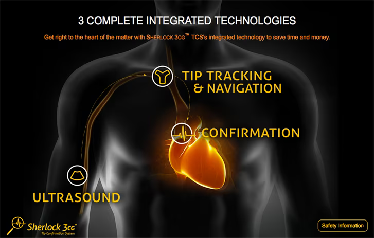
3cg-website.png
Discover the journey of a PICC

Clinical Study

Using X-ray to confirm PICC tip placement potentially delays time to treatment and exposes patients to harmful radiation.1

The Sherlock 3CG™ Tip Confirmation System (TCS) gets to the heart of the matter by using an adult patient's cardiac electrical activity to position the tip of the PICC in close proximity to the Cavo Atrial Junction (CAJ). Final tip location is confirmed by the nurse at the bedside, and infusion therapy can begin immediately without the need for a confirmatory chest x-ray.

Clinical Study Results

In a clinical study, 99.1% of 114 PICCs were placed at the CAJ or within +/- 1 cm using Sherlock 3CG™ Tip Confirmation technology.4

Hardware Options

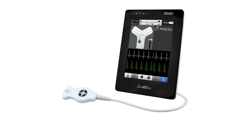
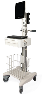
Sherlock 3CG™ Tip Confirmation System (TCS) simplifies the PICC insertion process by combining PICC tip placement technology into select Site~Rite™ Ultrasound systems.

The Site~Rite™ 8 Ultrasound System is currently available with integrated Sherlock 3CG™ Diamond TCS.

The Site~Rite™ 8 Ultrasound System with integrated Sherlock 3CG™ TCS is a "one-stop-shop" for PICC placements, allowing you these capabilities:

  • Evaluate and assess patient vasculature
  • Select and access the appropriate vessel
  • Use tip tracking technology to locate and navigate the catheter tip to the SVC
  • Use ECG technology to distinguish changes in P-wave amplitude and identify max P-wave
  • Confirm catheter tip placement at the Cavo Atrial Junction (CAJ) without the need for a confirmatory chest X-ray
  • DICOM® (Digital Imaging and Communications in Medicine) on select systems or printed documentation
EIFUs
Resources

References

Please consult labels and inserts for any indications, contraindications, hazards, warnings, cautions, and instructions for use.

  1. NICE guidelines 2015. “The Sherlock 3CG Tip Confirmation System for placement of peripherally inserted central catheters”.Chapter 6.1 https://www.nice.org.uk/guidance/mtg24
  2. Sanjay A. Patel et.al “Bedside Peripherally Inserted Central Catheter Tip Confirmation: A Direct Savings Analysis” JAVA.Vol 23 No 2.2018.
  3. Kenneth J Tomaszewski et.al “Time and resources of peripherally inserted central catheter insertion procedures: a comparison between blind insertion/chest X-ray and a real time tip navigation and confirmation system” ClinicoEconomics and Outcomes Research 2017:9 115–125.
  4. Data on file. BD, Salt Lake City, USA. 114 subjects evaluated; 113 confirmed. 

BD-58227

true